
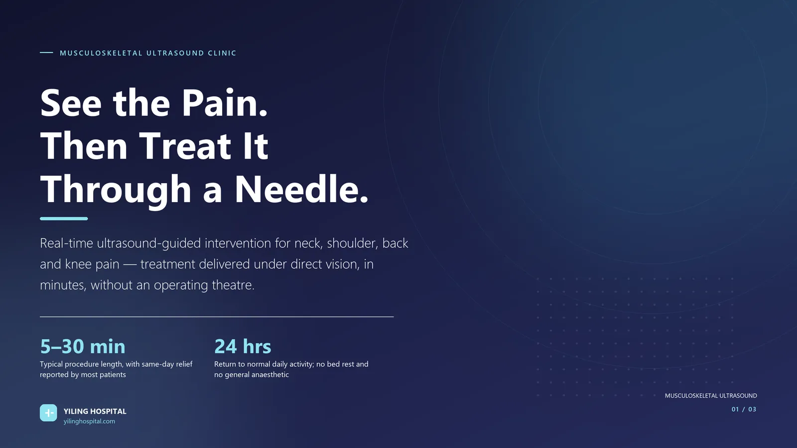
Musculoskeletal Ultrasound: Seeing the Pain, Then Treating It Through a Needle
Real-time ultrasound-guided intervention for neck, shoulder, back, knee and tendon pain — diagnosis and treatment in a single appointment.
Medically reviewed by the Musculoskeletal Ultrasound team, Yiling Hospital · Reading time approx. 10 minutes
A shoulder that cannot reach behind the back, a knee that swells after every flight of stairs, a wrist that aches when lifting a kettle, a heel that hurts on the first step out of bed. These are not vague complaints — each has a named anatomical structure that can be seen, measured and treated.
The problem is that they are rarely seen. An X-ray shows bone, not tendon. A physiotherapy assessment infers the lesion from movement. An injection given by surface landmark is, in effect, an educated guess: the medication may reach the inflamed bursa or it may stop two centimetres short of it, and nobody checks.
Musculoskeletal ultrasound changes that. It shows the tendon, the bursa, the joint lining and the nerve in real time, and — in the same visit, through the same probe — it guides a needle to exactly that structure. Diagnosis and treatment stop being two separate events separated by months.
What Musculoskeletal Ultrasound Shows That Other Imaging Does Not
Each imaging modality answers a different question, and choosing the wrong one is why patients accumulate reports that never explain the pain.
- X-ray — shows bone, joint space narrowing, fracture and calcification. It cannot see a tendon, a ligament or a bursa.
- MRI — shows everything anatomically, including bone marrow and deep soft tissue, and remains the gold standard for internal derangement and disc pathology. It is static, expensive, and often reported weeks after the scan.
- Ultrasound — shows soft tissue in motion: a tendon gliding through its sheath, a bursa compressing, a nerve thickening as it crosses a joint, a muscle contracting. It is dynamic, immediate, radiation-free, and repeatable.
The dynamic element matters more than it sounds. Many painful conditions are only visible when the joint moves — impingement that occurs at 70 degrees of abduction leaves no trace on a scan taken with the arm at rest. Ultrasound, held in the clinician’s hand while the patient moves, captures exactly the moment that reproduces the symptom.
Why Guidance Changes the Result
Landmark-guided injections rely on palpation and proportional measurements. Their accuracy varies widely and is frequently worse than assumed — the literature on shoulder and knee injections has repeatedly found that a meaningful proportion of blind injections do not reach the intended target.
Ultrasound guidance solves the accuracy problem directly: the needle is tracked continuously on screen, the target structure is confirmed before injection, and the spread of medication is watched in real time. Just as importantly, the structures that must not be hit — arteries, nerves, the pleura, the bowel — are identified and avoided before the needle advances rather than assumed to be somewhere else.
The most common reason a guided injection changes its plan mid-procedure is that something unexpected is found: a small artery in the needle path, a partial tear rather than inflammation, or a different structure entirely responsible for the pain. That is the value of looking.
The Advantages That Matter in Daily Life
Yiling Hospital’s musculoskeletal ultrasound clinic describes four core advantages, and each translates into something concrete for the patient.
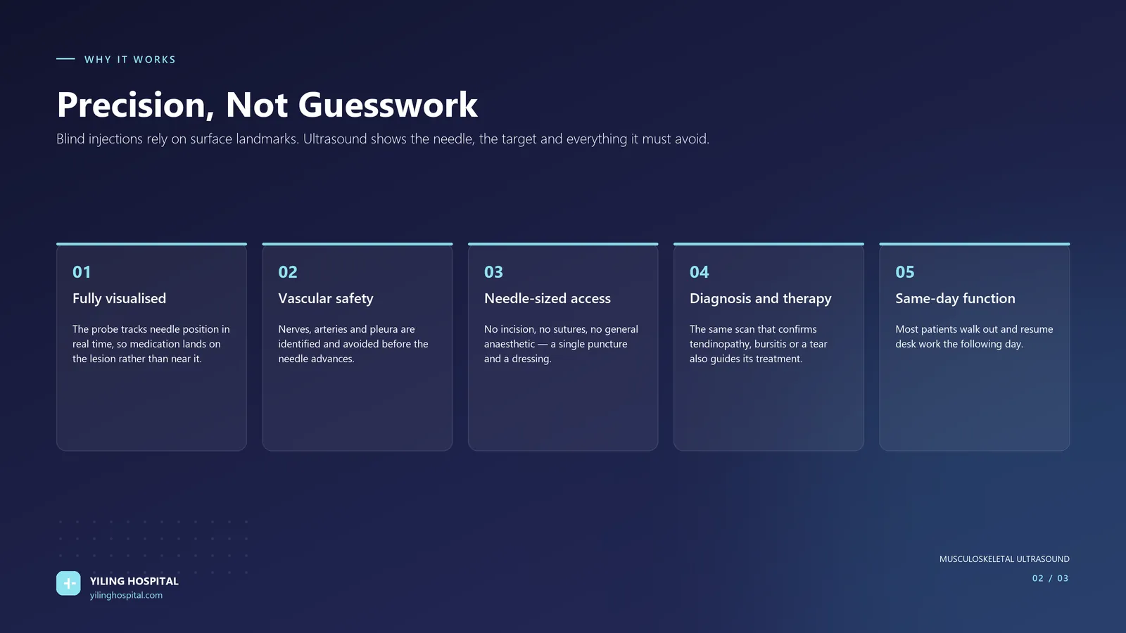
- Safe and minimally invasive — no incision, no sutures, no general anaesthetic. The wound is a needle puncture covered by a dressing.
- Fully visualised — the needle path and the target are both on screen throughout, so medication is delivered to the lesion rather than near it.
- Wide indication range — the same platform treats shoulder, cervical, lumbar, knee, wrist, elbow and foot pathology, and can be repeated if required.
- Fast — procedures typically take five to thirty minutes, and many patients report relief of pain the same day.
- Short recovery — no bed rest required; most patients return to normal daily activity within twenty-four hours, including desk work.
The Conditions This Clinic Treats
The clinic’s stated indications cover the musculoskeletal presentations that most often resist conservative care.
Table 1 — Common indications, what the scan shows and what the procedure does
| Condition | What ultrasound shows | What the guided procedure does |
|---|---|---|
| Frozen shoulder (adhesive capsulitis) | Thickened joint capsule, restricted movement, rotator interval changes | Hydrodilatation of the capsule plus targeted steroid, followed by a staged mobilisation programme |
| Rotator cuff tendinopathy and impingement | Tendon swelling, partial or full-thickness tear, subacromial bursitis | Subacromial or intratendinous injection placed under direct vision |
| Cervical spondylosis with neck stiffness and hand numbness | Nerve root calibre changes, facet joint changes, muscle spasm | Perineural or facet-targeted injection; release of myofascial trigger points |
| Lumbar disc herniation with radiating leg pain | Nerve root compression, surrounding inflammation | Epidural or perineural injection delivered to the compressed level |
| Knee osteoarthritis with swelling and stair difficulty | Cartilage thinning, effusion, synovitis, meniscal changes | Joint aspiration followed by hyaluronic acid or corticosteroid injection |
| Tenosynovitis — de Quervain’s, trigger finger | Thickened tendon sheath, fluid around the tendon, nodular thickening | Intra-sheath injection with the needle confirmed inside the sheath |
| Tennis elbow / golfer’s elbow | Common extensor or flexor origin tendinopathy, partial tear | Peppering technique or peritendinous injection at the origin |
| Plantar fasciitis and heel pain | Fascial thickening at the calcaneal insertion, plantar spur, bursitis | Fascial and bursal injection, with gait and footwear advice |
| Intercostal neuralgia | Intercostal nerve thickening, adjacent muscle spasm | Intercostal nerve block under direct vision |
| Lateral femoral cutaneous / superior cluneal nerve irritation | Nerve entrapment at known anatomical tunnels | Hydrodissection release around the nerve |
Frozen Shoulder: Where Guidance Makes the Biggest Difference
Adhesive capsulitis is the condition where blind treatment fails most visibly. The capsule contracts, external rotation is lost first, and the patient — often a 40–60-year-old with diabetes — spends eighteen months in pain with a shoulder that will not move.
A landmark-guided steroid injection may relieve pain temporarily but does nothing about the contracted capsule. Under ultrasound, two things become possible: accurate delivery of steroid into the rotator interval, and hydrodilatation — distending the capsule with fluid to mechanically stretch it. Combined with a staged mobilisation programme starting immediately afterwards, this addresses both the pain and the restriction.
Knee Osteoarthritis: Buying Function, Not Just Relief
Knee osteoarthritis is not one disease. Effusion-dominant, synovitis-dominant and mechanical patterns respond differently, and ultrasound distinguishes them in the clinic room. Where an effusion is present, aspirating it before injecting improves both comfort and drug delivery. Where synovitis is the driver, steroid is reasonable; where cartilage loss dominates with mechanical symptoms, hyaluronic acid or a different strategy altogether may be more appropriate.
The honest framing is that injection buys function and time. It does not regrow cartilage. Its value is in allowing a patient to complete the loading and strengthening programme that actually changes the trajectory of the disease — which is why the clinic pairs every procedure with an exercise plan.
Guided Injection Versus Physiotherapy, Steroids and Surgery
Patients rarely arrive at a clinic without having tried something. It is worth being clear about what each option does, because they are not interchangeable and they are not in competition.
Table 2 — Where each option fits
| Option | What it achieves | Best used when |
|---|---|---|
| Physiotherapy and loading programmes | Improves tissue capacity and tendon strength over weeks to months; the only option that changes what the tissue can tolerate | Always — as the foundation, and continued after any procedure |
| Landmark-guided injection | Delivers steroid near an assumed target without imaging | Now largely superseded where ultrasound is available; accuracy is variable |
| Ultrasound-guided injection | Confirms the diagnosis and delivers medication to the exact structure under direct vision | Pain that blocks rehabilitation; confirmed tendinopathy, bursitis, effusion, nerve entrapment |
| Hydrodissection | Separates a nerve or tendon from adherent surrounding tissue using fluid pressure | Nerve entrapment, adhesive scarring, restricted gliding |
| Surgery | Repairs a structural defect or releases a contracted structure | Full-thickness tears in appropriate candidates, mechanical block, failure of well-conducted conservative care |
The clinical logic is that an injection is a facilitator, not a treatment in itself. Its job is to reduce pain enough that the patient can do the loading work that actually changes the tissue. An injection given without a rehabilitation plan is a delay dressed as a cure.
What Happens During the Appointment
Where the indication allows, assessment and procedure happen in the same visit. That is deliberate: waiting weeks between diagnosis and treatment allows the acute inflammatory window to close and gives the patient time to lose confidence in the plan.
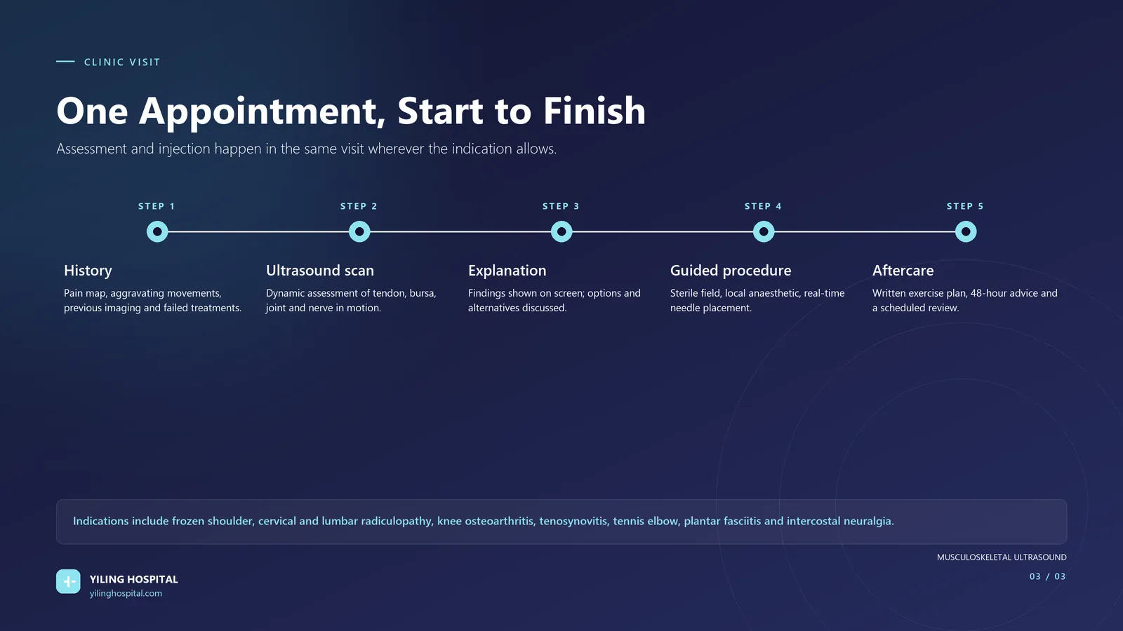
- History and examination — pain map, aggravating and relieving movements, previous imaging, and what has already been tried and failed.
- Ultrasound assessment — static and dynamic imaging of tendon, bursa, joint and nerve, with the patient moving the joint during the scan.
- Findings explained on screen — you see what the clinician sees, which is also why the treatment plan makes sense.
- Guided procedure — sterile field, local anaesthetic, real-time needle placement, confirmed spread of medication.
- Aftercare — written exercise plan, explicit 48-hour advice, and a scheduled review rather than ‘come back if it gets worse’.
Procedures take approximately 5–30 minutes. Most patients do not require bed rest and can resume normal daily activity within 24 hours. Any anticoagulant therapy should be declared before the appointment.
What to Expect Afterwards
Local anaesthetic wears off within a few hours, and a proportion of patients experience a temporary increase in soreness for 24–48 hours as the steroid crystals settle — this is expected and is not a sign that the procedure failed. The clinical effect of corticosteroid typically builds over three to ten days.
The most important aftercare instruction is not rest. It is controlled loading: the exercise programme begins within days, because the window in which pain is reduced is precisely the window in which strengthening is possible. An injection without rehabilitation is a temporary fix; an injection followed by a loading programme changes what the tissue can tolerate.
Frequently asked questions
The scan itself is not painful — it involves a probe on the skin with gel, though presssing on an inflamed structure can be tender. The guided procedure uses local anaesthetic, and most patients describe pressure rather than pain. The needle is fine (typically 23–25 gauge), and the injection itself takes under a minute.
A landmark-guided injection estimates the target from surface anatomy. An ultrasound-guided injection confirms it on screen, tracks the needle in real time and watches the medication spread. This improves accuracy, allows structures at risk to be avoided, and — critically — allows the diagnosis to be confirmed before treating. If the scan shows a different problem than expected, the plan changes.
Many patients need one. Some conditions — frozen shoulder, chronic tendinopathy, multi-level lumbar disease — require a short series or a repeat at a defined interval rather than repeated injections on demand. The clinic’s practice is to agree the number and timing in advance, because repeated steroid into a tendon without rehabilitation is a route to worse tissue quality, not better.
For most upper-limb and spinal procedures, yes — though it is sensible not to drive immediately after an injection into the shoulder or neck on the same day. Desk work can usually resume the next day. Heavy lifting and contact sport should wait until reviewed, typically one to two weeks depending on the site.
Diagnostic ultrasound uses no ionising radiation and is used routinely in pregnancy. The consideration is the injected medication rather than the imaging; corticosteroid and other injectables require individual discussion with the treating clinician, and some agents are avoided. Declare pregnancy or possible pregnancy before the appointment.
Then that is useful information, not a dead end. A normal scan redirects the search toward structures ultrasound cannot assess — bone marrow, deep joints, the spine itself — and toward non-structural contributors such as nerve sensitisation, sleep, and metabolic factors. The clinic refers onward for MRI or specialist assessment rather than proceeding with an injection that has no target.
The takeaway
Musculoskeletal pain is usually a mechanical problem with a specific anatomical cause. Musculoskeletal ultrasound identifies that cause in the clinic room and treats it through a needle in the same visit — with no incision, no general anaesthetic and, for most patients, a return to normal activity the next day. The procedure is quick; the difference is that someone finally looked.
Book a musculoskeletal ultrasound assessment
Bring any previous X-ray, MRI or ultrasound reports, and a list of current medication including blood thinners.
Yiling Hospital · Musculoskeletal Ultrasound Clinic · Shijiazhuang, Hebei, China
Medical disclaimer: this article is educational and does not constitute medical advice, diagnosis or a treatment recommendation. Suitability for any procedure can only be determined by a qualified clinician after examination. Individual recovery times and responses vary.
Department experts

Hand/foot microsurgery and ultrasound-guided needle-knife treatment for musculoskeletal pain.