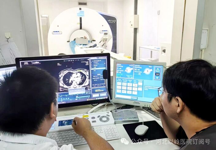
Precision imaging begins with advanced equipment. In today’s medical environment, patients seek higher diagnostic and treatment outcomes at lower medical costs. To provide the public with superior diagnostic services, Hebei Yiling Hospital recently introduced an ultra-high-end multi-slice spiral CT scanner. After installation and commissioning, it is now officially in use.
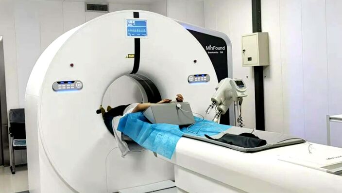
The newly introduced ultra-high-end CT features a domestically developed innovative true spherical wide-body detector, a world-leading magnetic levitation electromagnetic direct-drive system, and a fastest rotation speed of 0.25 seconds. It offers numerous advantages including faster scanning speed, higher image quality, lower radiation dose, and wider coverage, filling a technological gap in the ultra-high-end CT field.
80cm Large Aperture for Patient Comfort and Versatility
The spacious and comfortable scanning environment minimizes patient discomfort caused by confined spaces and alleviates anxiety, allowing patients to complete CT examinations in a natural and relaxed setting. It also facilitates easy scanning for obese patients or critically ill patients who need to carry medical devices.
True Spherical Shenguang Detector, Wide-Body and Versatile
The Shenguang detector uses Shengl Medical’s independently developed Shenguang material with a true spherical design. Compared to traditional detector materials, it has 50 times slower attenuation, twice the luminous efficiency, reducing radiation by 50%. Its afterglow time is reduced by 75% compared to traditional materials, minimizing interference between adjacent signal acquisitions. It improves sampling rate and data quality, ensuring excellent imaging quality while lowering radiation dose. During large-area whole-body scans, each detector pixel aligns perpendicularly to the X-ray tube focus, eliminating cone beam artifacts and achieving large-area, high-quality, low-dose scanning.
“Magnetic Levitation” Electromagnetic Direct Drive, Silent and Ultra-Fast
The Shenzhou gantry adopts a magnetic levitation electromagnetic direct-drive system, where the rotor and stator use electromagnetic direct drive technology with no friction or wear. It starts faster, with rotational accuracy far exceeding traditional belt-driven structures, and has a longer lifespan. This also significantly reduces noise during examinations, enhancing patient comfort throughout the scanning process.
NDI+ Micro-Dose Iterative Reconstruction Algorithm, Ultra-Low Dose
Utilizing an “intelligent multi-level dual-domain” iterative reconstruction—an AI-assisted physics-based iterative reconstruction algorithm—the original data undergoes simultaneous iteration in both projection and image domains. The projection domain iteration incorporates physical models of the X-ray tube and detector characteristics, while the image domain iteration employs a deep learning network based on anatomical structures to control iteration depth and restore image details at multiple scales. This enables ultra-low-dose CT examinations.
Advanced Intelligent Whole-Body Clinical Applications
This equipment achieves true free breathing, free heart rate, dynamic functional imaging of all organs, one-stop multifunctional imaging, whole-organ perfusion imaging, spectral imaging, and multimodal imaging. It transitions imaging from purely anatomical to functional, opening a new era in clinical CT applications.
Applications
One-Stop Chest Pain Examination: The ultra-high-end CT allows coronary CTA, pulmonary artery CTA, and aortic CTA to be completed simultaneously with a single contrast injection and scan. This technology provides a non-invasive, reliable method for the acute chest pain triple rule-out. One scan clearly displays the pulmonary artery, thoracic aorta, coronary arteries, and lung structures, providing multiple diagnostic insights and valuable time for identifying the exact cause and initiating emergency treatment.
One-Stop Stroke Examination: This includes non-contrast CT, CT perfusion (CTP), and CT angiography (CTA). Brain CTP has guiding clinical significance for stroke treatment strategies; CTA identifies culprit vessels related to stroke and assesses collateral circulation compensation.
One-Stop Ischemic Heart Disease Examination: With a single contrast injection, scan, and image reconstruction, this CT exam simultaneously obtains myocardial perfusion parameters, coronary artery images, and cardiac function analysis. It accurately evaluates coronary artery stenosis, myocardial blood supply, and the relationship between stenosis severity and myocardial perfusion.
Cardiac Coronary Artery Imaging: The gantry’s ultra-high rotation speed of 0.25 seconds completes coronary artery scans within a single cardiac cycle, achieving nearly 100% success rate. It is highly advantageous for screening coronary artery disease, and evaluating coronary stents and bypass grafts. Compared to traditional CT coronary CTA, this ultra-high-end spiral CT obtains clear images even in patients with high heart rates, arrhythmias, or atrial fibrillation, which is crucial for cardiovascular disease diagnosis and treatment.
Head and Neck Vascular Imaging Technology: For large-area imaging of head and neck vessels, it simultaneously observes the common carotid, internal carotid, external carotid, vertebral arteries, and intracranial arteries such as the anterior, middle, and posterior cerebral arteries. It detects aneurysms, plaque formation and characteristics, lumen stenosis and degree, and hypoperfusion. This enables early detection and precise localization of cerebral ischemia and infarction.
Whole-Body Large-Area Scanning: With wide coverage and rapid completion, various post-processing analysis software can display fine branches, clearly showing aneurysms, vascular malformations, stenosis or occlusions, atherosclerotic plaques, aortic dissection, tumor vasculature, and more.
Liver Multifunctional Imaging: Liver perfusion scanning automatically segments the liver, hepatic artery, hepatic vein, and portal vein. It supports automatic segmentation of the eight liver segments and manual segmentation based on portal vein/hepatic vein drainage. It features simulated surgical knife and puncture functions, calculates resected and residual liver volumes, enabling precise preoperative imaging assessment of intra-organ structures.
Spectral Multifunctional Imaging: Spectral CT overcomes the limitations of conventional CT in detecting iso-density lesions by generating images that directly reflect lesion tissue composition. It provides richer, more precise, and quantitative multiparametric imaging information. This is uniquely valuable for early tumor lesion detection, gout stone and calculus composition analysis, neurointerventional thrombectomy and thrombolysis contrast extravasation, and accurate differential diagnosis of cerebral hemorrhage.
Hebei Yiling Hospital has always adhered to a “patient-centred” service philosophy. In recent years, it has continuously increased investment in equipment upgrades, quality enhancement, and professional refinement. The introduction of this ultra-high-end multi-slice spiral CT will further elevate the hospital’s diagnostic and treatment capabilities, providing the public with more convenient, rapid, precise, and safe medical imaging services, ensuring patients receive timely, effective, and high-quality medical care.
— Hebei News Network, 23 August 2024